Aqueous Extract of Azadirachcta indica Protect Against Gentamicin-Induced Hepatotoxicity via Regulation of Lipid Peroxidation, Oxidative Stress and Inflammation but Enhances Apoptotic Markers
-
Oluwadare Joshua Ogundipe


Department of Human Physiology, Faculty of Basic Medical Sciences, Redeemer’s University, Ede, Osun, Nigeria
Abodunrin Adebayo OjetolaDepartment of Physiology, Faculty of Basic Medical Sciences, Adeleke University, Ede, Osun, Nigeria
Omolola Funke AkinpeluDepartment of Human Anatomy, Faculty of Basic Medical Sciences, Olabisi Onabanjo University, Ago-Iwoye, Ogun, Nigeria
Olaoluwa Sesan OlukiranDepartment of Physiological Sciences, Faculty of Basic Medical Sciences, Obafemi Awolowo University, Ile-Ife, Nigeria
Rufus Ojo AkomolafeDepartment of Physiological Sciences, Faculty of Basic Medical Sciences, Obafemi Awolowo University, Ile-Ife, Nigeria
Adetomiwa Ezekiel AdeogunDepartment of Physiology, Faculty of Basic Medical Sciences, Ladoke Akintola University of Technology, Ogbomoso, Oyo, Nigeria
| Received 10 Nov, 2023 |
Accepted 20 Feb, 2024 |
Published 01 Jan, 2025 |
Background and Objective: Gentamicin administration caused oxidative stress in the liver, damaging the lipids, proteins and DNA in liver cells by producing reactive oxygen species. Thus, the possible effect of the aqueous extract of Azadirachta indica (AAI) on gentamicin-induced liver injury was investigated. Materials and Methods: The study involves twenty adult male Wistar rats (housed in 4 separate plastic cages) such that a graded dosage of AAI was administered to the experimental group for 14 days (p.o.) before exposure to gentamicin toxicity (100 mg/kg) for one week. At the end of the study, comparisons of some markers of liver enzymes, antioxidant status and Inflammatory and apoptotic markers were made between the control, GEN and AAI-pretreated groups at p<0.05. Results: The gentamicin treatment caused a significant increase (p<0.05) in AST, ALP, Bilirubin, MDA, Catalase, Interleukin-1, Caspase-3, Bcl-2 as well as a significant decrease (p<0.05) in TNFα, BAX and SOD level. Pre-treatment with graded doses of AAI caused a significant increase in ALP, MDA, SOD, GPx, Bcl-2 and BAX, as well as a significant decrease (p<0.05) in AST, Bilirubin, CAT, Interlukin-1 and Caspase-3. There was an appreciable difference in the histoarchitecture of the liver of AAI pre-treated groups compared to the GEN. Conclusion: Hence, the extract has prophylactic potential against gentamicin-induced liver injury, although a risk profile of liver apoptosis is not unlikely on 200 mg AAI pre-treatment, as evidenced by the increase in Bcl-2 and BAX.
INTRODUCTION
The liver is an essential organ that carries out numerous tasks, such as immunity, digestion, detoxification, metabolism and vitamin storage1. Gentamicin, a regularly used antibiotic from the aminoglycoside class, can potentially harm the Liver in addition to its well-known nephrotoxicity2. The specific liver damage from Gentamicin involves various processes and may alter liver function. Examining the mechanism of action and potential adverse effects of gentamicin is essential to understanding the precise nature of gentamicin-induced liver damage. Gentamicin administration can cause oxidative stress in the liver, damaging the lipids, proteins and DNA in liver cells by producing reactive oxygen species (ROS). Gentamicin can also change mitochondrial membrane potential, decrease oxidative phosphorylation and switch mitochondrial permeability, all of which can harm and malfunction cells. Additionally, gentamicin use can result in the release of pro-inflammatory cytokines like Interleukin-1 Beta (IL-1 beta), Tumor Necrosis Factor-Alpha (TNF-α) and Interleukin-6 (IL-6), which can lead to hepatocellular inflammation and tissue damage2.
Neem (Azadirachta indica) has a long history of conventional use. It has long been prized for its healing abilities throughout many cultures. Native to the Indian subcontinent, neem is now grown all over the world. It has bioactive substances, including Nimbin, nimbidin, azadirachtin and gedunin, that support its therapeutic effects3. Neem has long been used to treat fever, infections, skin conditions and digestive issues. Its leaves, bark, seeds and oil are all utilized medicinally in different ways. Neem has been used for centuries and contemporary scientific study has confirmed its use and identified new uses, such as for dental health, wound healing, immune system regulation and insect-repellent characteristics4. Azadirachta indica has antiviral, antibacterial, antifungal and anti-inflammatory properties; despite its usefulness, there is a gap in the literature on the use of Azadirachta indica on gentamicin-induced liver injury; hence, this study focused on the effects of aqueous extract of Azadirachta indica on gentamicin-induced liver injury.
MATERIALS AND METHODS
Study area: This study was done at the Animal House of the Faculty of Basic Medical Sciences, Department of Human Physiology Redeemer’s University Ede, Osun State, Nigeria. The duration of this study was 5 weeks; 2 weeks for acclimatization and 3 weeks for the administration of extracts.
Chemicals and drugs: Normal saline, phosphate buffer, formalin, distilled water (gotten from Redeemer’s University Biochemistry Laboratory), ten packs of Gentamicin injection 80 mg/2 mL (manufactured by Shanxi Shuguang Pharmaceutical Co. Ltd.) marketed as antibiotic drugs active against a wide variety of pathogenic Gram-negative and positive bacteria.
Plant extracts sensitivity test
Collection and identification of plants: The leaves of the plants (Azadirachta indica) were sourced from their farms across South West, Nigeria, with the help of traditional herb sellers. Dr. Nidrea George, a Botanist at the Department of Botany, University of Lagos, identified Azadirachta indica.
Preparation of plant materials: The plants were prepared according to the method of Thapa and Wilson5. The leaves were washed thoroughly under tap water and shade-dried at room temperature (24-26°C). An electric grinder pulverized the dried leaves. The powder was passed through a 40 mesh sieve and stored in an air-tight container before use.
Plants extraction method: The aqueous extract was prepared using the cold maceration soxhlet extraction techniques6. Five grams of each plant extract were weighed into 100 mL of extraction solvents (water and ethanol) and left on a mechanical shaker (The Corning LSE™ Orbital Shaker) overnight at room temperature. The extract solutions were filtered aseptically into another 100 mL reagent bottle using a Wattman No. 1 filter paper. All the filtrate was screened by inoculation unto nutrient agar plates and incubated at 37°C for 48 hrs Orhue and Nwanze7 to make sure that the extracts were sterile for further analysis.
Phytochemical analysis of plant extracts: Leaf extracts were evaluated by a qualitative phytochemical analysis6 to determine their major constituents. The presence of phytochemicals was analyzed by following standard procedures, which are as follows:
• |
Test for tannins: The test for tannins was carried out by subjecting 3 g of each plant extract to 6 mL of distilled water, filtered and ferric chloride reagents added to the filtrate |
|
• |
Test for basic alkaloids (Mayer’s Test)5: Five millimeters of extract were concentrated to yield a residue. The residue was dissolved in 3 mL of 2% (v/v) HCl. Three drops of Mayer’s reagents were added. The appearance of the dull white precipitate indicated the presence of primary alkaloids |
|
• |
Test for saponins5: Two millimeters extracts were shaken vigorously for 30 sec in a test tube. After 30 min of shaking, the presence of thick forth will indicate saponins |
|
• |
Test for terpenoids5: One millimeter of the extract was mixed with 2 mL of chloroform. Three millimeters of concentrated H2SO4 were added to form a layer. Reddish brown precipitate coloration at the interface indicated the presence of terpenoids |
Animal care and management: Twenty adult male Wistar rats weighing 100-120 g were used in this study; they were obtained from the Animal House of Redeemers University, Ede and were housed in plastic cages. The animals were kept under normal environmental conditions with a 12 hrs light/dark cycle and had free access to a standard rodent pellet diet (Top Feeds P.L.C. Osogbo, Nigeria) and water. They were allowed to acclimatize in the laboratory for two weeks before the commencement of the study. The Faculty of Basic Medical Sciences Ethical Review Committee (FBMSERC) at Redeemers University in Ede, Osun State, granted ethical clearance. By FBMSERC recommendations, the rats received humane treatment.
Experimental design: The rats were divided into four groups: Group 1 (the control), consisting of 5 rats, received distilled water daily orally for six weeks. Group 2 through 4 received different treatments. Five rats from Group 2 (the toxic control) were given 100 mg/kg/day of Gentamicin via intraperitoneal (i.p.) for a week (Table 1). Twenty four hours after the last gentamicin injection, five rats were slaughtered and treated similarly to the Group 1 animals. There were five rats in Group 3 and 4. They got gentamicin after receiving pre-treatments of 100 and 200 mg/kg/day per oral (p.o.) of AAI, respectively, for 14 days. Five rats from each group were sacrificed by cervical dislocation 24 hrs after injection. These rats’ blood was drawn from their hearts and placed in individual EDTA bottles. A cold centrifuge was used to separate the plasma, which was then tested for liver enzymes, antioxidant markers and inflammatory and apoptotic markers. For histological research, the livers of each of these rats were removed, weighed and preserved in 10% formalin. The body weights of the rats were assessed once a week during the study period.
Descriptive and inferential statistics were used to analyze the collected data.
Measurement of body weight: A digital weighing scale (Camry weighing balance, China) was used to measure the body weight of the rats once a week during the study period to track weight changes in each group.
| Table 1: | Representing the study design | |||
| Groups | 1-7 days | 8-14 days | 14-21 days |
| Group 1 | DW | ||
| Group 2 | GEN (100 mg/kg) | ||
| Group 3 | AAI (100 mg/kg) | AAI (100 mg/kg) | GEN (100 mg/kg) |
| Group 4 | AAI (200 mg/kg) | AAI (200 mg/kg) | GEN (100 mg/kg) |
| DW: Distilled water, GEN: Gentamicin, AAI: Aqueous extract of Azadirachta indica, Group 1: Control, Group 2: GEN, Group 3: 100 mg/kg AAI+GEN and Group 4: 200 mg/kg AAI+GEN | |||
Biochemical assay: The cold centrifuge (Centurium Scientific, Model 8881, UK) was used to centrifuge 5 mL of whole blood collected into a heparinized tube for 30 min at 5000 rpm. The indicators of liver function, such as AST, ALP, γ-GGT and Bilirubin, were examined in the plasma after it had been separated.
Assessment of antioxidant status: The liver was carefully excised, weighed and homogenized with 10 mL of sucrose solution (0.25 M) using an Electric Homogenizer (SI601001). The homogenate was centrifuged at 3000 rpm for 20 min and the supernatant was collected for the assessment of malondialdehyde (MDA) levels, reduced Glutathione (GSH) glutathione peroxidase (GPx) and superoxide dismutase (SOD).
Assessment of markers of inflammations and apoptosis: The supernatant collected from the liver homogenate was assessed for Interleukin, Caspase-3, Tumor Necrotic Factor-Alpha (TNF-α), Bcl-2 Associated X (BAX) and B-Cell Lymphoma 2 (Bcl-2).
Histopathological examination: The liver was carefully excised, weighed and fixed in 10% formo-saline solution. Thereafter, the tissues were embedded in paraffin wax, sectioned and stained using hematoxylin and eosin.
Ethical clearance: All experiments were undertaken according to regulations set out by the Redeemers University Health Research and Ethical Committee (RUHREC) with Reference number (RUN/REC/2023/061).
Statistical analysis: GraphPad Prism-5 was the statistical package used for data analysis. One-way Analysis of Variance (ANOVA) was used to analyze data, followed by the Students Newman-Keuls (SNK) test for multiple comparisons. Results were expressed as Mean±SEM and p<0.05 was taken as the accepted level of significant difference.
RESULTS
Body weight of rats at the initial: There was a significant difference in body weight of Group 2 and 4 compared to the control (F = 39.28, p<0.0001), while a decrease in Group 3 and 4 compared to Group 2 as shown in Table 2.
Body weight of rats at week 1: There was a significant difference in body weight of Group 2, 3 and 4 compared to the control (F = 38.12, p<0.0001), while a decrease in Group 3 and 4 compared to Group 2 as shown in Table 2.
Body weight of rats at week 2: A significant decrease was observed in Group 3 and 4 compared to Group 2 (F = 8.452, p = 0.0001) as shown in Table 2.
Body weight of rats at week 3: There was a significant decrease in body weight of Group 2 and 3 compared to the control (F = 33.81, p<0.0001), while an increase in Group 3 and 4 compared to Group 2 and also in Group 4 compared to Group 3 as shown in Table 2.
| Table 2: | Effects of aqueous extract of Azadirachta indica on body weight (g) of rats with gentamicin-induced hepatotoxicity | |||
| Body weight | Control | GEN | 100+GEN | 200+GEN |
| Initial weight | 113.6±1.40 | 137.0±1.38α | 105.0±2.83β | 98.20±0.80αβ |
| Week 1 | 134.6±1.29 | 160.2±2.99α | 119.0±2.05αβ | 123.40±1.29αβ |
| Week 2 | 158.0±2.17 | 169.3±6.17 | 146.8±4.77β | 152.00±2.12β |
| Week 3 | 185.6±3.87 | 160.0±2.39α | 174.6±4.85αβ | 186.80±2.73βδ |
| Values are expressed in Mean±SEM (n = 5) p<0.05, αp<0.05 vs control, βp<0.05 vs GEN, δp<0.05 vs 100+GEN | ||||
|
| Table 3: | Effects of aqueous extract of Azadirachta indica on enzymes parameters of rats with gentamicin-induced hepatotoxicity | |||
| Parameter | Control | GEN | 100+GEN | 200+GEN |
| γ-GGT (U/L) | 3.05±0.275 | 4.08±0.741 | 4.34±0.289 | 3.76±0.620 |
| ALT (U/L) | 9.15±0.119 | 11.25±0.75α | 9.50±0.204β | 9.33±0.24β |
| AST (U/L) | 6.61±0.478 | 8.72±0.196α | 5.11±0.723β | 3.99±0.420αβ |
| ALP (U/L) | 2.71±0.050 | 9.42±0.613α | 24.75±1.495αβ | 14.92±1.784αβδ |
| Bilirubin (mg/dL) | 1.38±0.042 | 1.69±0.10α | 1.02±0.03αβ | 1.02±0.04αβ |
| Values are expressed in Mean±SEM (n = 5), p<0.05, αp<0.05 vs control, βp<0.05 vs GEN and δp<0.05 vs 100+GEN | ||||
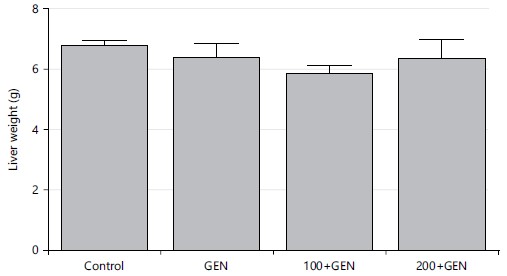
Liver weight change: There was no significant difference in liver weight of Group 2, 3 and 4 (6.40±0.47, 5.87±0.25 and 6.34±0.65) when compared with Group 1 (6.80±0.15) (p = 0.0666, F = 2.479) as shown in Fig. 1.
Gamma-Glutamyl Transferase (γ-GGT) (U/L) concentration in plasma: There was no significant difference in the plasma concentration of γ-GGT of Group 2, 3 and 4 when compared with Group 1 (p = 0.3740, F = 1.135) as shown in Table 3.
Alanine Transaminase (ALT) concentration in plasma: There was a significant increase in the plasma concentration of ALT of Group 2 when compared with group 1 (p = 0.012, F = 5.619), while a significant decrease in Groups 3 and 4 compared to group 2 as shown in Table 3.
Aspartate Transaminase (AST) concentration in plasma: There was a significant difference in plasma concentration of AST of Group 2 and 4 rats when compared with Group 1. Also, a significant decrease in Group 3 and 4 when compared to Group 2 (p = 0.0001, F = 17.33) as shown in Table 3.
Alkaline Phosphatase (ALP) concentration in plasma: The plasma concentration of ALP in Group 2, 3 and 4 rats was significantly higher (p≤0.0001, F = 59.92) when compared with Group 1, also group 3 and 4 when compared to Group 2 and 3 when compared to Group 4 as shown in Table 3.
Bilirubin concentration in plasma: There was a significant difference in the plasma concentration of bilirubin of Group 2, 3 and 4 when compared with group 1, also in Group 3 and 4 when compared with Group 2, (p≤0.0001, F = 29.15) as shown in Table 3.
|
| Table 4: | Effects of aqueous extract of Azadirachta indica on antioxidant parameters of rats with gentamicin-induced hepatotoxicity | |||
| Parameter | Control | GEN | 100+GEN | 200+GEN |
| MDA (μM/g) | 0.66±0.119 | 1.92±0.131α | 1.86±0.059α | 2.62±0.128αβδ |
| GSH (μ/mg) | 0.03±0.002 | 0.023±0.0001α | 0.029±0.001β | 0.030±0.002β |
| SOD (μ/mg) | 2.25±0.250 | 1.00±0.204α | 2.50±0.354β | 6.00±0.456αβδ |
| CAT (μ/mg) | 2.53±0.295 | 7.32±0.269α | 1.03±0.015αβ | 2.28±0.254βδ |
| GPx (μ/mg) | 1.64±0.008 | 1.74±0.05 | 3.74±0.11αβ | 6.98±0.740αβδ |
| Values are expressed in Mean±SEM (n = 5) p<0.05, αp<0.05 vs control, βp<0.05 vs GEN and δp<0.05 vs 100+GEN | ||||
Malonaldehyde (MDA) concentration in liver: The MDA concentrations in Group 2, 3 and 4 rats were significantly higher (p≤0.0001, F = 51.57) when compared with Group 1, also Group 4 when compared to Group 2 and 3 as shown in Table 4.
Glutathione (GSH) concentration in liver: There was a significant decrease in GSH concentrations in Group 2 when compared with group 1 (p = 0.045, F = 3.616). while a significant increase in Group 3 and 4 compared to Group 2 as shown in Table 4.
Superoxide dismutase (SOD) concentration in liver: There were significant differences in SOD concentrations of Group 2 and 4 when compared with group 1 (p≤0.0001, F = 42.05), while a significant increase in Group 3 compared with Group 2 as shown in Table 4.
Catalase (CAT) concentration in liver: The catalase concentration in Group 2 was significantly higher (p≤0.0001, F = 135.7) when compared with Group 1, while Group 3 and 4 were lower when compared to Group 2. Also, an increase was recorded in Group 4 compared to Group 3 as shown in Table 4.
Glutathione Peroxidase (GPx) concentration in liver: The GPx concentrations in Group 3 and 4 were significantly higher (p≤0.0001, F = 49.97) when compared with Groups 1 and 2, also Group 4 when compared with Group 3 as shown in Table 4.
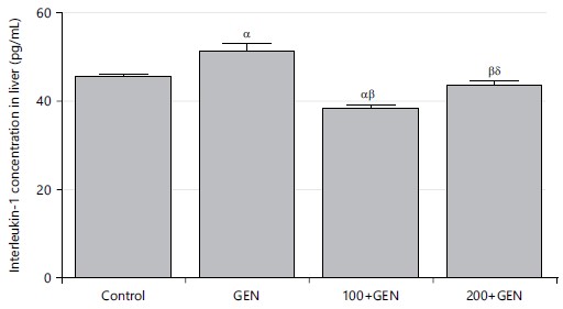
Interleukin-1 concentration in liver: There was a significant difference in interleukin-1 concentration of Group 2 and 3 (51.45±1.517 and 38.33±0.923) when compared with Group 1 (45.65±0.378) (p≤0.0001, F = 27.42), while a significant decrease in Group 3 and 4 (38.33±0.923 and 43.55±1.001) compared to Group 2 (51.45±1.517). Also an increase in Group 4 (43.55±1.001) when compared to Group 3 (38.33±0.923) as shown in Fig. 2.
|
|
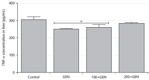
Tumor Necrotic Factor-Alpha (TNF-α) concentration in liver: There was a significant difference in TNF-α concentration of Group 2 and 3 (251.40±2.135 and 259.90±16.81) when compared with Group 1 (305.60±15.57) (p = 0.0309, F = 4.161) as shown in Fig. 3.
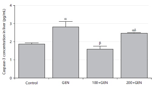
Caspase-3 concentration in liver: The caspase-3 concentrations in Group 2 and 4 (2.80±0.322 and 2.46±0.047) were significantly higher (p = 0.0024, F = 8.705) when compared with Group 1 (1.88±0.049), while Group 3 (1.58±0.179) was lower when compared with Group 2 (2.80±0.322). Also, an increase was recorded in Group 4 (2.46±0.047) when compared with Group 3 (1.58±0.179) as shown in Fig. 4.
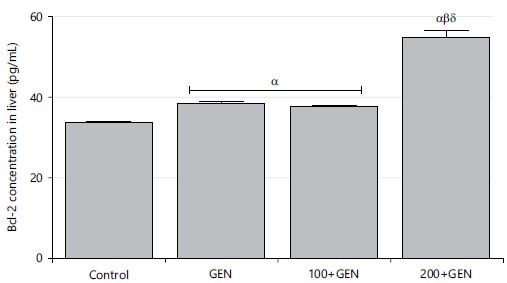
Bcl-2 concentration in the liver: The Bcl-2 concentration in Group 2, 3 and 4 (38.50±0.500, 37.76±0.238 and 55.00±1.732) was significantly higher (p≤0.0001, F = 105.0) when compared with the Group 1 (33.60±0.294), also, an increase was recorded in Group 4 (55.00±1.732) when compared to Group 2 and 3 (38.50±0.500 and 37.76±0.238) as shown in Fig. 5.
|
|
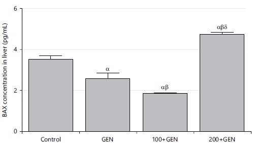
BAX concentration in liver: The BAX concentrations in Group 2 and 3 (2.56±0.279 and 1.86±0.027) were significantly lower (p≤0.0001, F = 49.30) when compared with the Group 1 (3.52±0.182), also, a decrease was recorded in Group 3 (1.86±0.027) when compared with Group 2 (2.56±0.279). In contrast, an increase was recorded in Group 4 (4.71±0.111) compared to Group 3 (1.86±0.027) as shown in Fig. 6.
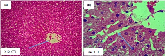
Histopathological examination: The Control group Plate 1 shows normal liver tissue, no disruption in the sinusoidal cells (long blue arrow), no abnormal vacuolation and no fibrotic lesions were present.
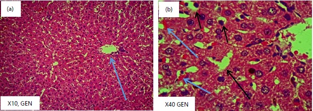
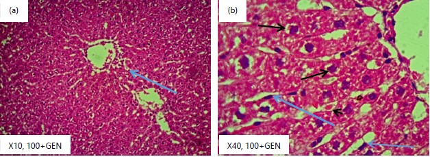
The GEN group Plate 2 shows Sinusoidal cell disruption, (long black arrow), vacuolation of the hepatic cell (short black arrows) and numerous fibrotic lesion (long blue arrows).
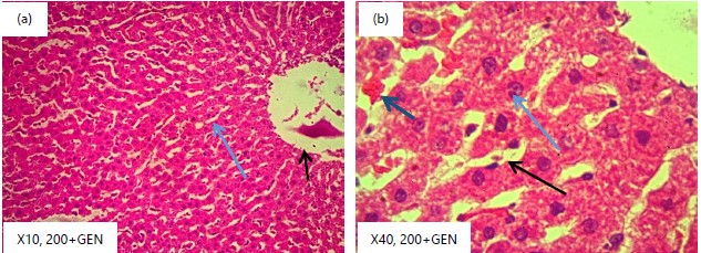
The Group C Plate 3 rats show reduced fibrotic lesions, (short black arrow), mild vacuolation of the hepatic cell (long black arrow) and normal sinusoidal cells (long blue arrow).
The Group D Plate 4 rats show Fewer fibrotic lesions (Thick blue arrow) but normal hepatic and sinusoidal cell (slender and black arrows).
|
|
|
|
DISCUSSION
This study investigates the effect of the aqueous extract of Azadirachta indica on some markers of hepatic function, antioxidant markers, inflammatory and apoptotic markers of rats with gentamicin-induced liver injury.
The decrease in body weight as observed in this study Table 2 is an indication that gentamicin harms the body system resulting in weight loss. This was similar to the findings of El-Zawahry and Abu El Kheir8, who reported that gentamicin administration caused a reduction in food intake and body weight of rats. Weight loss is one of the known effects of Gentamicin on body weight. According to El-Zawahry and Abu El Kheir8, using Gentamicin for an extended period or at a high dosage may cause weight loss. The drug’s tendency to harm the kidneys and subsequently impair renal function is thought to be the leading cause of this weight loss9. According to Lopez-Novoa et al.10 Gentamicin belongs to the class of antibiotics known as aminoglycosides, which have nephrotoxic (destructive to the kidney) effects. Cohen et al.11 stated that the kidneys preserve the body’s fluid and electrolyte equilibrium, increase urine and the loss of vital minerals like potassium and calcium can result from changes in managing fluid and electrolytes when the kidneys are damaged. In addition, gentamicin-induced renal impairment can cause decreased appetite and food malabsorption, leading to further weight loss12. Individuals may occasionally experience gastrointestinal issues, including nausea and vomiting, which can affect their ability to eat and as a result, loss of body weight13. The advantages of using gentamicin to treat a severe bacterial infection often outweigh any adverse effects, including weight loss14 overall, even though gentamicin can result in weight loss as a side effect (Table 2).
No significant increase in liver weight was recorded in all the experimental groups compared to the control group as shown in Fig. 1. This was in contrast to the findings of Hassan et al.15 and Saeed et al.16, who reported that Gentamicin may increase liver weight, which could be a sign of hepatomegaly or an enlarged Liver. Gentamicin-treated experimental animal models frequently show this increase in liver weight16. Gentamicin’s hepatotoxic effects can harm the liver, resulting in inflammation and cellular changes in the liver tissue, which help explain the observed rise in liver weight. In addition, oxidative stress and disruption of liver cellular processes are likely to be the mechanisms driving gentamicin’s hepatotoxicity15. The nephrotoxic effects of the antibiotic may also subtly affect liver function, adding to changes in liver weight.
The breakdown of glutathione is facilitated when γ-GGT increases, this leads to its depletion and causes DNA and structural protein damage induced by oxidative stress. The elevated level of γ-GGT in plasma experimental groups compared to the control group. Table 3 agreed with the findings of Lushchak17, who reported that the level of γ-GGT in the hepatocytes is elevated with an increase in oxidative stress. Gentamicin may also have a hepatoxic effect, increasing the γ-GGT levels in the plasma, which suggests liver dysfunction18. Group 3 and 4 show that Azadirachta indica at higher doses barely restores the γ-GGT level toward the control. Furthermore, the study’s results show that pre-treatment with Azadirachta indica at higher doses restores the rat liver to normal function19.
There was a significant increase in the plasma concentration of ALT in Gentamicin group compared to the control group Table 3. This was in agreement with the findings of Smith et al.14. High levels of ALT in the blood can indicate liver dysfunction. Gentamicin has been reported to increase serum ALT, indicating possible hepatotoxic effects14.
Although Azadirachta indica leaves have hepatoprotective effects, Table 3 shows that Azadirachta indica has positive effects. This agreed with Mishra et al.20, who investigated the hepatoprotective effects of neem leaf extract in rats with liver damage induced by carbon tetrachloride. The study results showed that neem leaf extract significantly reduced the levels of ALT in the treated rats, suggesting a potentially beneficial effect on liver function20.
A significant increase in the concentration of AST in plasma was observed in the gentamicin rats when compared with the control group Table 3 and the increase also indicates liver injury, which is similar to the findings of Yoon et al.21 that studied hepatotoxic effects of Gentamicin in rats. The results showed that gentamicin administration led to a significant increase in AST levels in the treated rats, suggestive of liver damage. Azadirachta indica showed hepatoprotective ability which is in similarity with Ojha et al.22. The findings demonstrated that treatment with neem leaf extract significantly reduced the elevated levels of AST in the CCl4-induced liver injury rats, suggesting a protective effect on liver function.
A significant increase in the concentration of ALP in plasma was observed in the gentamicin rats compared with the control group Table 3, this was similar to the reports of Ibraheem et al.23. The increased ALP levels in the rats are an indication of liver injury. Azadirachta indica has no preventive effects on the harmful impact of gentamicin on the liver. This was in contrast with Ojewole et al.24, which showed that neem extract significantly reduced the elevated levels of ALP in the rats, indicating a potential protective effect on the liver.
There was a significant increase in the bilirubin concentration, which was observed in the gentamicin-treated rats when compared with the control group Table 3; the increase could be due to the harmful effects of gentamicin on the liver, leading to its inability to process bilirubin due to bile duct obstruction; this is similar to the findings of Ogundipe et al.25, who reported that gentamicin causes increase in bilirubin level which also suggest damage on liver function.
The MDA (Malondialdehyde) is a marker to detect lipid peroxidation and oxidative stress. The MDA concentration in Group 2 is significantly higher than in the control group Table 4. This indicated that gentamicin administration has induced oxidative stress and lipid peroxidation in the liver, leading to cellular damage and potential liver injury. The MDA concentration in Group 3 is also significantly higher than in the control group but lower than in Group 2. This suggested that Azadirachta indica treatment may have some protective effect on the liver by reducing oxidative stress and lipid peroxidation to some extent.
Glutathione (GSH) is a naturally occurring antioxidant found in the cells of plants, animals and many microorganisms. The decrease in the glutathione level in the group two rats is evidence of increased catabolism of glutathione as evident in the increased level of γ-GGT which connotes the presence of oxidative stress in the liver. Group 3 and 4 show that Azadirachta indica restores the glutathione level toward the control Table 4. From this study, pre-treatment with Azadirachta indica protects the liver from oxidative damage.
Superoxide dismutase (SOD) is an essential antioxidant enzyme that protects cells from oxidative stress. The significant decrease in SOD concentration in Group 2 suggests that the liver is under oxidative stress Table 4. With lower levels of SOD, the liver’s ability to neutralize superoxide radicals is compromised, accumulating damaging ROS26. This oxidative damage can harm liver cells, disrupt normal liver functions and contribute to the development of liver injury or dysfunction. The significant increase in SOD in Group 3 compared to Group 2 suggests that Azadirachta indica administration helped to restore SOD levels, potentially mitigating the oxidative stress induced by gentamicin. The increased SOD may aid in reducing the harmful effects of ROS and protect liver cells from further damage. The significant increase in SOD in Group 4 compared to the control group and Group 2 suggests that the protective capacity of Azadirachta indica is dose-dependent. This increase may offer substantial protection against oxidative stress, potentially reducing liver damage and promoting liver cell recovery27.
Glutathione peroxidase (GPx) is an essential enzyme found in organisms, including humans, that plays a crucial role in protecting cells from oxidative damage caused by reactive oxygen species (ROS) or free radicals28. The significant increase in GPx concentration in Group 3 and 4 indicated that the administration of Azadirachta indica protects the Liver against the oxidative stress induced by gentamicin Table 4. Azadirachta indica or neem, is known for its potent antioxidant and anti-inflammatory properties, which likely contribute to this protective effect29. The higher GPx concentration in Group 4 compared to Group 3 further supports the idea that Azadirachta indica supplementation has a dose-dependent effect on enhancing the antioxidant defense system in the liver. This means a higher dose of Azadirachta indica provides better protection against gentamicin-induced oxidative stress.
A significant increase in the concentration of interlukin-1 in plasma was observed in the gentamicin-treated rats when compared with the control group Fig. 2; the increase could be as gentamicin triggering the inflammatory process. These observed changes are to the report of El-Barbary et al.30. Similar to renal disease, gentamicin use has been linked to liver damage, which can trigger inflammatory processes and cause the release of pro-inflammatory cytokines like IL-1. The reverse was the case in the level of TNF-α in the liver tissue of gentamicin treated rats as shown in Fig. 3.
The effects of gentamicin on liver function and its influence on cytokine levels, especially IL-1, have been documented in several investigations. For instance, a 2015 study examined the hepatotoxic effects of gentamicin in rats. It was published in the Journal of Applied Toxicology. The presence of inflammation and liver damage was indicated by the researchers’ observation of an increase in IL-1 levels in the liver tissue of gentamicin-treated rats30. Epoxyazadiradione has anti-inflammatory properties against macrophage migration inhibitory factors31. Azadirachtin suppresses retinoic acid-mediated biological reactions32.
Caspase-3 is a critical protein involved in the process of apoptosis. In Group 2, the significantly higher caspase-3 concentration indicates a substantial increase in apoptosis compared to the control group Fig. 4. This suggested that gentamicin treatment is causing cellular damage and initiating a significant apoptotic response in the liver, which could be detrimental to liver health. In Group 3, the caspase-3 concentration is lower than in Group 2. This finding suggests that the administration of neem may have a protective effect on the liver. The lower caspase-3 levels imply that neem treatment might mitigate the apoptotic response triggered by Group 2, potentially reducing cellular damage. In Group 3, the caspase-3 concentration is higher than in the control but lower than in Group 2 Fig. 4. This finding suggested that neem alone might be causing a mild increase in apoptosis, which could be a normal response to the treatment but not the extent of causing severe cellular damage.
The BAX is a member of the Bcl-2 family of proteins and it falls under the pro-apoptotic group, which means it promotes cell death. The Bax concentration is significantly lower (Fig. 6) while the Bcl-2 increases in gentamicin-induced rats compared to the control group. This finding suggested that gentamicin-induced liver injury is associated with a decrease in Bax and an increase in Bcl-2 expression. This decrease in Bax and increased expression of Bcl-2 might be related to a potential cellular defense mechanism against the damaging effects of gentamicin or it could indicate a shift in the balance of pro-apoptotic and anti-apoptotic signals in favor of cell survival. In Group 3, the Bax concentration is significantly lower (Fig. 6) and increased Bcl-2 level Fig. 5 compared to both the control group and Group 2. This suggested that neem treatment is further reducing Bax expression compared to gentamicin-induced liver injury alone. The downregulation of Bax and upregulation of Bcl-2 may indicate that neem offers protective effects against gentamicin-induced liver injury by potentially inhibiting or minimizing the apoptotic response.
Several researchers33-35 show that in experimental models of liver injury brought on by chemicals like carbon tetrachloride (CCl4), paracetamol and ethanol, it has been demonstrated that neem extracts and compounds display hepatoprotective properties. These studies have shown that neem can lessen oxidative stress, enhance liver function markers such as serum liver enzyme levels and histological alterations and lessen liver injury.
CONCLUSION
It is evident that gentamicin causes hepatotoxicity despite its powerful action against gram-negative and positive bacteria, also neem leaf extract administration before gentamicin treatment was able to protect the liver from gentamicin toxic effects. Thus, the aqueous extract of Azadirachcta indica protects gentamicin-iduced hepatotoxicity via regulation of lipid peroxidation, oxidative stress and inflammation but enhances apoptotic markers.
SIGNIFICANCE STATEMENT
Gentamicin is one of the most potent antibiotics used against gram-positive and negative bacteria and its use is limited due to harmful effects on some organs such as the liver and kidney. It has been reported that Azadirhacta indica has antibacterial, anti-inflammatory and antiviral properties. Despite its usefulness, there is a gap in the literature on the use of this plant against the harmful effects of gentamicin on the liver.
REFERENCES
- Sherman, C.B., S. Behr, J.L. Dodge, J.P. Roberts, F.Y. Yao and N. Mehta, 2019. Distinguishing tumor from bland portal vein thrombus in liver transplant candidates with hepatocellular carcinoma: The A-VENA criteria. Liver Transplant., 25: 207-216.
- Kane-Gill, S.L., 2021. Nephrotoxin stewardship. Crit. Care Clin., 37: 303-320.
- Yadav, D.K., Y.P. Bharitkar, K. Chatterjee, M. Ghosh, N.B. Mondal and S. Swarnakar, 2016. Importance of neem leaf: An insight into its role in combating diseases. Indian J. Exp. Biol., 54: 708-718.
- Biswas, K., I. Chattopadhyay, R.K. Banerjee and U. Bandyopadhyay, 2002. Biological activities and medicinal properties of neem (Azadirachta indica). Curr. Sci., 82: 1336-1345.
- Thapa, R. and G.D. Wilson, 2016. The importance of CD44 as a stem cell biomarker and therapeutic target in cancer. Stem Cells Int., 2016.
- Bamidele, O., D.S. Arokoyo, A.M. Akinnuga and A.O. Oluwarole, 2014. Antidiabetic effect of aqueous extract of Basella alba leaves and metformin in alloxan-induced diabetic albino rats. Afr. J. Biotechnol., 13: 2455-2458.
- Orhue, N.E.J. and E.A.C. Nwanze, 2004. Effect of Scoparia dulcis on Trypanosoma brucei induced alterations in serum transaminase, alkaline phosphatase and bilirubin in the rabbit. J. Med. Sci., 4: 194-197.
- El-Zawahry, B.H. and E.M. Abu El Kheir, 2007. The protective effect of curcumin against gentamicin-induced renal dysfunction and oxidative stress in male albino rats. Egypt. J. Hosp. Med., 29: 546-556.
- Hayward, R.S., J. Harding, R. Molloy, L. Land, K. Longcroft-Neal, D. Moore and J.D.C. Ross, 2018. Adverse effects of a single dose of gentamicin in adults: A systematic review. Br. J. Clin. Pharmacol., 84: 223-238.
- Lopez-Novoa, J.M., Y. Quiros, L. Vicente, A.I. Morales and F.J. Lopez-Hernandez, 2011. New insights into the mechanism of aminoglycoside nephrotoxicity: An integrative point of view. Kidney Int., 79: 33-45.
- Cohen, L., R. Lapkin and G.J. Kaloyanides, 1975. Effect of gentamicin on renal function in the rat. J. Pharmacol. Exp. Ther., 193: 264-273.
- Miller, G., H. Schaefer, S. Yoder, R. Miller and P. Winokur et al., 2018. A randomized, placebo-controlled phase I trial of live, attenuated herpes zoster vaccine in subjects with end-stage renal disease immunized prior to renal transplantation. Transplant Infect. Dis., 20.
- Jackson, S.E., L. Holter and R.J. Beeken, 2019. ‘Just because I’m old it doesn’t mean I have to be fat’: A qualitative study exploring older adults’ views and experiences of weight management. BMJ Open, 9.
- Smith, H.L., A.K. Berglund, J.B. Robertson, L.V. Schnabel, R.J. McMullen Jr., B.C. Gilger and A. Oh, 2023. Effect of gentamicin on CD3+ T-lymphocyte proliferation for treatment of equine recurrent uveitis: An in vitro study. Vet. Ophthalmol., 26: 347-354.
- Hassan, S.M., M.M. Khalaf, S.A. Sadek and A.M. Abo-Youssef, 2017. Protective effects of apigenin and myricetin against cisplatin-induced nephrotoxicity in mice. Pharm. Biol., 55: 766-774.
- Saeed, N.M., E. El-Demerdash, H.M. Abdel-Rahman, M.M. Algandaby, F.A. Al-Abbasi and A.B. Abdel-Naim, 2012. Anti-inflammatory activity of methyl palmitate and ethyl palmitate in different experimental rat models. Toxicol. Appl. Pharmacol., 264: 84-93.
- Lushchak, V.I., 2012. Glutathione homeostasis and functions: Potential targets for medical interventions. J. Amino Acids, 2012.
- Babaeenezhad, E., N. Nouryazdan, M. Nasri, H. Ahmadvand and M.M. Sarabi, 2021. Cinnamic acid ameliorate gentamicin-induced liver dysfunctions and nephrotoxicity in rats through induction of antioxidant activities. Heliyon, 7.
- Baligar, N.S., R.H. Aladakatti, M. Ahmed and M.B. Hiremath, 2014. Hepatoprotective activity of the neem-based constituent azadirachtin-A in carbon tetrachloride intoxicated wistar rats. Can. J. Physiol. Pharm., 92: 267-277.
- Mishra, R.K., R. Saini, D.J.P. Kumar, R. Sankannavar, P. Binnal, N. Dwivedi and P. Kumar, 2023. Thermo-catalytic pyrolysis of Azadirachta indica seeds over CaO and CuO: Pyrolysis kinetics, impact of catalysts on yield, fuel properties and its chemical compositions. J. Energy Inst., 111.
- Yoon, E.J., Y.N. Chabane, S. Goussard, E. Snesrud, P. Courvalin, E. Dé and C. Grillot-Courvalin, 2015. Contribution of resistance-nodulation-cell division efflux systems to antibiotic resistance and biofilm formation in Acinetobacter baumannii. Biom, 6.
- Ojha, S., B. Venkataraman, A. Kurdi, E. Mahgoub, B. Sadek and M. Rajesh, 2016. Plant-derived agents for counteracting cisplatin-induced nephrotoxicity.Oxid. Med. Cell. Longevity, 2016.
- Ibraheem, Z.O., S.S. Farhan, A. Al Sumaidaee, L. Al Sufi, A. Bashir, A. Balwa and R. Basir, 2021. Liver functions in combined models of the gentamicin induced nephrotoxicity and metabolic syndrome induced by high fat or fructose diets: A comparative study. Toxicol. Res., 37: 221-235.
- Ojewole, E., I. Mackraj, P. Naidoo and T. Govender, 2008. Exploring the use of novel drug delivery systems for antiretroviral drugs. Eur. J. Pharm. Biopharm., 70: 697-710.
- Ogundipe, O.J., O.F. Akinpelu, A. Oyerinde and O.R. Oluwakemi, 2021. Ocimum gratissimum (Linn) leaves extract attenuates oxidative stress and liver injury in gentamicin-induced hepatotoxicity in rats. Egypt. J. Basic Appl. Sci., 8: 146-155.
- Gill, S.S. and N. Tuteja, 2010. Reactive oxygen species and antioxidant machinery in abiotic stress tolerance in crop plants. Plant Physiol. Biochem., 48: 909-930.
- Mehta, A. and S.J.S. Flora, 2001. Possible role of metal redistribution, hepatotoxicity and oxidative stress in chelating agents induced hepatic and renal metallothionein in rats. Food Chem. Toxicol., 39: 1029-1038.
- Antunes, F., D. Han and E. Cadenas, 2002. Relative contributions of heart mitochondria glutathione peroxidase and catalase to H2O2 detoxification in in vivo conditions. Free Radical Biol. Med., 33: 1260-1267.
- Lokanadhan, S., P. Muthukrishnan and S. Jeyaraman, 2012. Neem products and their agricultural applications. J. Biopesticides, 5: 72-76.
- El-Barbary, M.N., R.I.H. Ismail and A.A.A. Ibrahim, 2015. Gentamicin extended interval regimen and ototoxicity in neonates. Int. J. Pediatr. Otorhinolaryngology, 79: 1294-1298.
- Alam, M.R., M.R. Amin, A.K.M.A. Kabir, M. Moniruzzaman and D.M. McNeill, 2007. Effect of tannins in Acacia nilotica, Albizia procera and Sesbania acculeata foliage determined in vitro, in sacco, and in vivo. Asian Australas. J. Anim. Sci., 20: 220-228.
- Thoh, M., B. Babajan, P.B. Ragavendra, C. Sureshkumar and S.K. Manna, 2011. Azadirachtin interacts with retinoic acid receptors and inhibits retinoic acid-mediated biological responses. J. Biol. Chem., 286: 4690-4702.
- Bhanwra, S., J. Singh and P. Khosla, 2000. Effect of Azadirachta ondica (Neem) leaf aqueous extract on paracetamol-induced liver damage in rats. Indian J. Physiol. Pharmacol., 44: 64-68.
- Baligar, N.S., R.H. Aladakatti, M. Ahmed and M.B. Hiremath, 2014. Evaluation of acute toxicity of neem active constituent, nimbolide and its hepatoprotective activity against acute dose of carbon tetrachloride treated albino rats. Int. J. Pharm. Sci. Res., 5: 3455-3466.
- Baliga, M.S., A.R. Shivashankara, S. Venkatesh, H.P. Bhat, P.L. Palatty, G. Bhandari and S. Rao, 2019. Phytochemicals in the Prevention of Ethanol-Induced Hepatotoxicity: A Revisit. In: Dietary Interventions in Liver Disease: Foods, Nutrients, and Dietary Supplements, Watson, R.R. and V.R. Preedy (Eds.), Academic Press, Cambridge, Massachusetts, ISBN: 9780128144664, pp: 79-89.
How to Cite this paper?
APA-7 Style
Ogundipe,
O.J., Ojetola,
A.A., Akinpelu,
O.F., Olukiran,
O.S., Akomolafe,
R.O., Adeogun,
A.E. (2025). Aqueous Extract of Azadirachcta indica Protect Against Gentamicin-Induced Hepatotoxicity via Regulation of Lipid Peroxidation, Oxidative Stress and Inflammation but Enhances Apoptotic Markers. Science Digest, 1(1), 1-15. https://doi.org/10.21124/sd.2025.01.15
ACS Style
Ogundipe,
O.J.; Ojetola,
A.A.; Akinpelu,
O.F.; Olukiran,
O.S.; Akomolafe,
R.O.; Adeogun,
A.E. Aqueous Extract of Azadirachcta indica Protect Against Gentamicin-Induced Hepatotoxicity via Regulation of Lipid Peroxidation, Oxidative Stress and Inflammation but Enhances Apoptotic Markers. Science Digest 2025, 1, 1-15. https://doi.org/10.21124/sd.2025.01.15
AMA Style
Ogundipe
OJ, Ojetola
AA, Akinpelu
OF, Olukiran
OS, Akomolafe
RO, Adeogun
AE. Aqueous Extract of Azadirachcta indica Protect Against Gentamicin-Induced Hepatotoxicity via Regulation of Lipid Peroxidation, Oxidative Stress and Inflammation but Enhances Apoptotic Markers. Science Digest. 2025; 1(1): 1-15. https://doi.org/10.21124/sd.2025.01.15
Chicago/Turabian Style
Ogundipe, Oluwadare, Joshua, Abodunrin Adebayo Ojetola, Omolola Funke Akinpelu, Olaoluwa Sesan Olukiran, Rufus Ojo Akomolafe, and Adetomiwa Ezekiel Adeogun.
2025. "Aqueous Extract of Azadirachcta indica Protect Against Gentamicin-Induced Hepatotoxicity via Regulation of Lipid Peroxidation, Oxidative Stress and Inflammation but Enhances Apoptotic Markers" Science Digest 1, no. 1: 1-15. https://doi.org/10.21124/sd.2025.01.15

This work is licensed under a Creative Commons Attribution 4.0 International License.